View Cervical Spine Anatomy Diagram Background. The cervical spinal nerves, sometimes called nerve rootlets , exit the spinal canal through the neuroforamen in pairs—1 nerve exits on the left side and 1 on the right. Anatomy of the human spine complete with illustrations and references.
Anatomy Of The Spine Southern California Orthopedic Institute from www.scoi.com
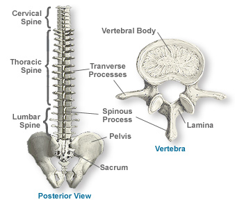
The cervical spine is the most superior portion of the vertebral column, lying between the cranium and the thoracic vertebrae. Middle and lower cervical spine. International anatomical terminology by the federative committee on anatomical terminology (fcat).
The cervical spinal nerves, sometimes called nerve rootlets , exit the spinal canal through the neuroforamen in pairs—1 nerve exits on the left side and 1 on the right.
The cervical spine becomes a lordotic curve, like the lumbar spine. It runs from the neck to the upper back. The cervical spine (neck) starts at the base of skull and extends down to the thoracic spine. It moves the head around and has a lot of freedom to move in all directions.